Narrowing the Immunotherapy Window Through Use of a Programmable Cellular Interface
Authors: Colleen M. O’Connor, Andres Campos, Elizabeth Goodson, Aurora Fabry-Wood, Jinchun Wang, Andries Zijlstra
StimulusBio at the International Society for Cell & Gene Therapy (ISCT) on May 7-10, 2025
in New Orleans, LA
Download the Full ISCT Poster Here
Background:
- The Immune Synapse is a membrane complex of co-stimulatory molecules (co-stim), antigens, and other factors organized into a higher order structure.
- Current cell expansion technologies can’t replicate this organization or mediate selective cell expansion.
- Programming molecular mediators of immune activity is necessary to selectively expand specific immune phenotypes.
- Red Blood Cells (RBCs) provide a biocompatible framework to present costim molecules in a controlled manner.
Rationale:
- To advance CAGT innovation and clinical impact, we developed a customizable Programmable Cellular Interface (sbPCI) on an RBC platform with proof of concept demonstrated initially on sbPCI-1.
Methods:
- Autologous and allogeneic RBCs were modified to display an array of sbPCI-1 molecular configurations.
- PBMCs from 3 donors were co-cultured at a 1:10 ratio (sbPCI-1:PBMC ) and exogenous IL-2 (50U/ml) & IL-21 (30ng/ml) added 3x per week.
- Lymphocytes were re-stimulated and analyzed every 8 days.
- Total RNA was extracted from all 3 donor PBMCs, plus corresponding expanded T cells at Days 8 and 24.
- cDNA was synthesized for qPCR use with TaqMan gene expression assays.
- Error bars are Standard Error of the Mean (SEM).
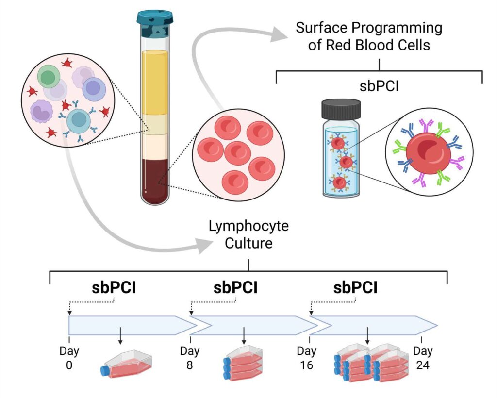
Figure 1: Programmable Cellular Interface Platform. RBCs were isolated from donor blood and modified to display sbPCI consisting of co-stim molecules. Lymphocytes were then expanded on sbPCI.
Results:
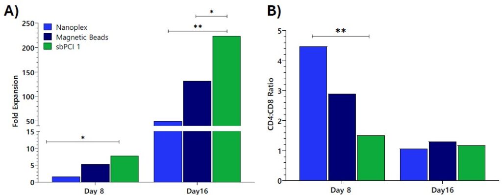
Figure 2: Performance Comparison: sbPCI-1 vs. Leading Competitors. PBMCs were expanded, according to manufacturer’s directions, and compared with sbPCI-1. The sbPCI-1 did not require the initial T-cell isolation step. A) sbPCI-1 had improved fold expansion over 16 days. B) sbPCI-1 showed similar performance, with a decrease in CD4:CD8 ratios observed over a period of 16 days. P-values calculated using a two-way ANOVA (*p < 0.05, **p < 0.01) suggested differences observed were significant. These observations supported further development of the sbPCI system.

Figure 3: Modular Design for Rapid Modification of the Molecular Formats. A) Molecular configurations can be easily optimized with the sbPCI system to drive desired outcomes. B) Unstimulated PBMCs co-cultured 48 hours with sbPCI, harvested and then analyzed for Granzyme B expression by flow cytometry. C) Cells were enumerated over 24 days to determine total cell expansion. D) 24-day fold expansion was calculated based on cell numbers with sbPCI-1 iii showing a reduced fold expansion by Day 24.
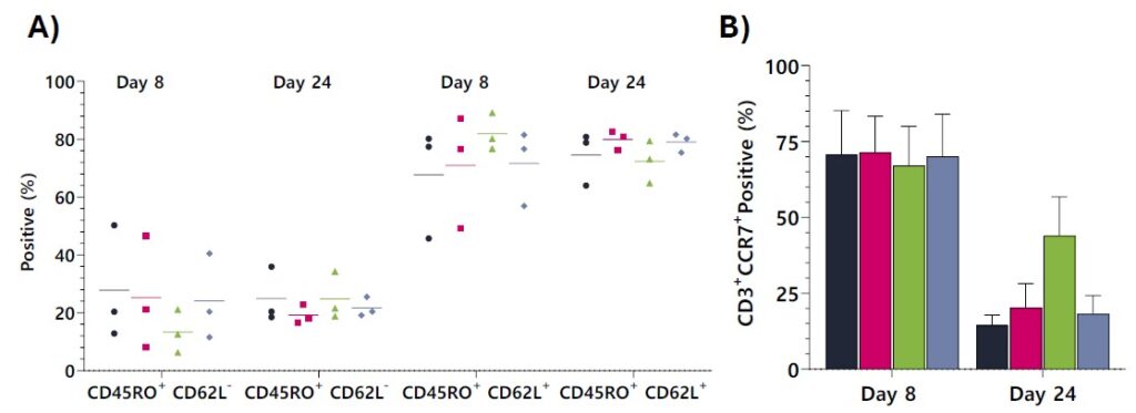
Figure 4: Molecular Formats Selectively Expand Central Memory (CM) T-cells vs Effector Memory (EM) T-cells. PBMCs were co-cultured with sbPCI over 24 days and analyzed by flow cytometry and gene expression every 8 days. A) All sbPCI formats produced primarily CM T-cells by Day 8 and maintained this phenotype. B) At Day 8, the majority of T cells were positive for CCR7+. By Day 24, there was an increased number of CD3+CCR7+ expressing cells in the sbPCI-1 iii cultures compared with other sbPCI-1 configurations.

Figure 5: Molecular Formats Increase CD3+CD8+. A) Day 24 expanded cells’ phenotype was evaluated by flow cytometry and expression measured as the percent positive. B) The sbPC-1 iii expanded a majority of CD3+CD8+.
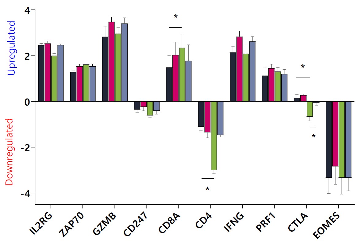
Figure 6: T-cell Activation Pathways Are Upregulated on Day 24. mRNA was analyzed for log2 fold changes between formats, with unpaired t-tests determining significance (*p < 0.05). CTLA-4 expression was dependent on the sbPCI-1 format.
Conclusion:
- The molecular configuration of co-stim molecules presented to PBMCs can be easily optimized using the sbPCI system.
- Changes in molecular formats affect T-cell expansion and phenotype.
- Expansion ranged from 1,115x to 2,025x, without the need for T-cell isolation.
- sbPCI-1 iii expanded primarily CD8+ Tcells, suggesting the CD3+CD8+ isolation step could be removed.
- sbPCI-1 supported CM T-cells expansion.
- CTLA-4 expression was influenced by the sbPCI-1 molecular format.
Future Directions:
- These findings warrant further experiments testing types, placement, and concentrations of different co-stim molecules and antigen combinations.
- Future sbPCI molecular formats aim to enhance cell therapy precision by focusing on: CAR-T, TCR-specific, NK/NKT cells, and iPSCs.
- Collaborations with manufacturing sites to demonstrate patient-specific use.
Contact StimulusBio to Talk About Your Scientific and Commercial Product Needs
In the competitive landscape of life science tools and diagnostics, even groundbreaking innovations struggle to gain traction and achieve widespread adoption. At StimulusBio, we specialize in empowering these vital technologies to reach their full potential. With our proven commercialization strategies and deep industry expertise, we partner with you at every stage to optimize your product’s marketability, reliability, and profitability – setting you up for lasting success in the marketplace.

